False Profile hip
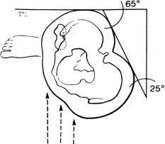
First, position the patient 90 degrees to the image receptor
Second, rotate the pelvis and feet from 90 degrees to form an approximately 65 degrees with the image receptor.
Image source:www.orthopaedia.com
Second, rotate the pelvis and feet from 90 degrees to form an approximately 65 degrees with the image receptor.
Image source:www.orthopaedia.com
|
False Profile Hip
*Important positioning notes *The femur of the affected side should be perpendicular to the floor. *The patient should NOT be leaning and the knee should be locked on the affected side. *The foot of affected side should be parallel to the image receptor. *The foot of unaffected side should be abducted and/or perpendicular to the image receptor. *The degree in obliquity will vary from patient to patient. Centering: the central ray should exit the hip of interest. Stand behind the x-ray tube to assess centering. Too much collimation light seen lateral to the affected side will indicate off centering (too much soft tissue). Film size: 8x10 or 10x12 in Radiation protection: Gonad shielding is a contraindication in males and females since it will obscure the anatomy. Criteria A false profile hip projection demonstrates anterior acetabular coverage of the femoral head . A false profile allows measurement of the anterior center-edge and shows evidence anterior subluxation while weight bearing. A properly positioned false profile hip image will demonstrate the "bullet-sign". The "bullet-sign" is the product of the superimposed ischial tuberosity of the affected side. |
|
Right false profile hip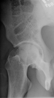
Right false profile hip image properly demonstrating the "bullet sign".
Left false profile hip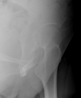
Left false profile hip image properly demonstrating the "bullet sign".
|
Right false profile hip positioning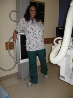
Right profile hip positioning. Note abduction of unaffected leg and affected side is perpendicular with foot parallel to image receptor.
Left false profile hip positioning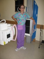
Left false profile hip positioning. Note alternative positioning of feet at right angles. The pelvis is correctly obliqued to demonstrate the "bullet sign".
|
Right false profile hip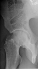
Right false profile hip
|
Right false profile hip positioning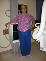
The affected leg should be perpendicular and the knee should be locked to ensure weight-bearing. The affected foot should be parallel to the image receptor and the unaffected foot should be perpendicular to the image receptor.
|
