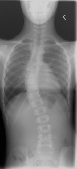
Scoliosis
- PA (reduces the dose to the gonads by 10% of the entrance dose
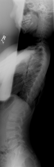
- PA & LAT of the entire spine standing (unless otherwise requested)
- Films should include entire spine from the level of E.A.M. to the ASIS at a 72-inch SID on a 34" cassette
- Collimation should allow inclusion of pelvic crests and rib cage.
- DO NOT use breast shields on a first time PA scoliosis projection.
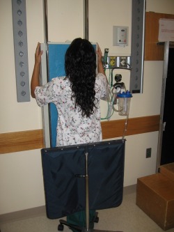
- Patient should stand with weight evenly distributed, feet apart to keep balance.
- The patient should not lean forward or backward.
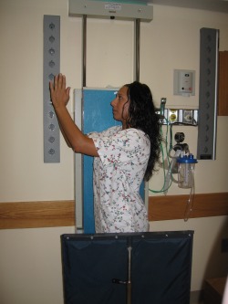
- The arms should be in the "benediction" position. (The humeri should be approximately 30 degrees to the coronal plane with the palms of the hands facing each other in the sagittal plane.)
- The midsagittal plane of the skull and the torso should be perpendicular to the image plate.
- The body should be against the image plate and chin elevated. The identical position should be used for the lateral. The purpose is to keep from rotating the spine and/or shoulders.
- The top of the image plate should be at the EAM in order to place the majority of the anatomy on the top portion of the image plate.The display algorithm for the stitched image is calculated based on this placement.
- Use the portashield at the level of the A.S.I.S.
- Use breast shields on follow-up exams.
- If breast shields cannot be used on the lateral projection, do a left-lateral on an even day and a right-lateral on an odd day since this will divide the dose among the breast tissue closest to the x-ray tube.
- When measuring the patient to determine the technical factors, place the calipers at the level of the breast for a male and just under the breast for a female. Close the calipers tightly and take the inner-line measurement.
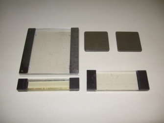
- Filtering for PA projection: Place the bottom of the PA filter at the level of T-7.
- Filtering for the Lateral projection: Place the bottom of the cervical filter at the top of the shoulders.
 |
 |
 |
 |
 |
 |
